Featured Publications

AI-guided Prospective Cancer Radiotherapy

We describe an AI model that generates radiation treatment plans for prostate cancer patients. AI was successfully deployed in a standard-of-care clinical setting, providing gains in efficiency, improved treatment quality, and an understanding of physicians’ decision-making and perceptions of AI use when patient care is at stake.

Improving Safety and Model Interpretability in Radiomics

Refinement of radiomic results and methodologies is required to ensure progression of the field. In this work, we establish a set of safeguards designed to improve and support current radiomic methodologies through detailed analysis of a radiomic signature.

First Voxel-Based AI for Radiation Therapy Planning

We present a proof-of-concept for a method to automatically infer the radiation dose directly from the patient’s treatment planning image based on a database of previous patients with corresponding clinical treatment plans. Our method uses regression forests augmented with density estimation over the most informative features to learn an automatic atlas-selection metric that is tailored to dose prediction.

Predicting contour quality

Our method uses random forests to learn joint distributions over the training features, and then exploits a set of learned potential group configurations to build a conditional random field (CRF) that ensures the assignment of labels is consistent across the group of segmentations. The CRF is then solved via a constrained assignment problem. We validate our method on 1574 plans, consisting of 17579 segmentations, demonstrating an overall classification accuracy of 91.58%.

Image segmentation on a learned manifold

We highlight the effects of varying the parameters and initialization on segmentation accuracy and propose a framework for attaining improved results using image adaptive parameters and initializations through novel applications of manifold learning.

  • August 01, 2023

    Aly Khalifa and Yuan Gao are awarded CIHR CGS-D fellowships for 3-years!

  • August 01, 2023

    Sangwook Kim awarded DSI fellowship for 3-years!

  • May 11, 2023

    Sangwook Kim successfully passes his PhD reclassification exam on Multi-Task Learning For Robust Deep Learning-Based Medical Image Analysis!

  • May 11, 2023

    Yuan Gao successfully passes his PhD reclassification exam on Temporal Deep Learning Applications in Healthcare Monitoring and Wearables!

  • July 8, 2022

    Aly Khalifa successfully passes his PhD reclassification exam on Automated Treatment Planning for Adaptive Radiation Therapy!

  • June 29, 2021

    Siham successfully passes her PhD reclassification exam on Deep learning for Automatic Medical Image Segmentation!

  • May 28, 2021

    Cathy successfully passes her PhD reclassification exam on Developing an AI Image-Based Biomarker for Interventional Cardiovascular Risk-Stratification!

  • June 01, 2019

    McIntosh Lab secures PMCC Innovation Award for proposal of an AI-driven system for automatic coronary angiographic interpretation.

Director

Chris McIntosh

  • Chair in Medical Imaging and Artificial Intelligence
  • Scientist: Techna Institute, Peter Munk Cardiac Centre, Joint Department of Medical Imaging, University Health Network
  • Assistant Professor: Department of Medical Biophysics, Department of Computer Science, University of Toronto
  • Faculty Affiliate, Vector Institute
  • Contact information
  • Chris McIntosh is a Computer Scientist, trained in Computer Vision and AI in medicine. He is the recipient of academic awards from NSERC, CIHR, and the Michael Smith Foundation for Health Research. His PhD dissertation developed AI-driven methods for quantitative medical image analysis through manifold learning, and received an honourable mention for the top dissertation in computer vision and medical image analysis by the Canadian Image Processing and Pattern Recognition Society.

    His research focuses on the theory and clinical application of AI in medicine for improving patient care including transfer learning, meta learning, computer vision, and explainable AI. Applications include deep learning for automated diagnosis, segmentation, quality assurance, and treatment planning. His past work on AI in radiation therapy has been approved for clinical use by regulatory bodies, commercialized, and deployed in hospitals around the world, using AI to deliver reproducible, high quality cancer care.

    Outside of work Chris is a father, golfer, and a snowboarder.

Students and Staff

Siham Amara-Belgadi (PhD Student)

William Gao (PhD Student)

Sangwook Kim (PhD Student)

Sejin Kim (MSc Student)

Cathy Ongly (PhD Student)

Balagopal Unnikrishnan (PhD Student)

Aly Khalifa (MSc Student)

Overview

Working together

Innovation doesn't happen in a vacuum. Jointly embedded in Canada's largest hospital network and the University of Toronto, our research brings together experts and trainees in computer science, medicine, biology, and physics to solve impactful problems at the intersection of artificial intelligence and medicine.

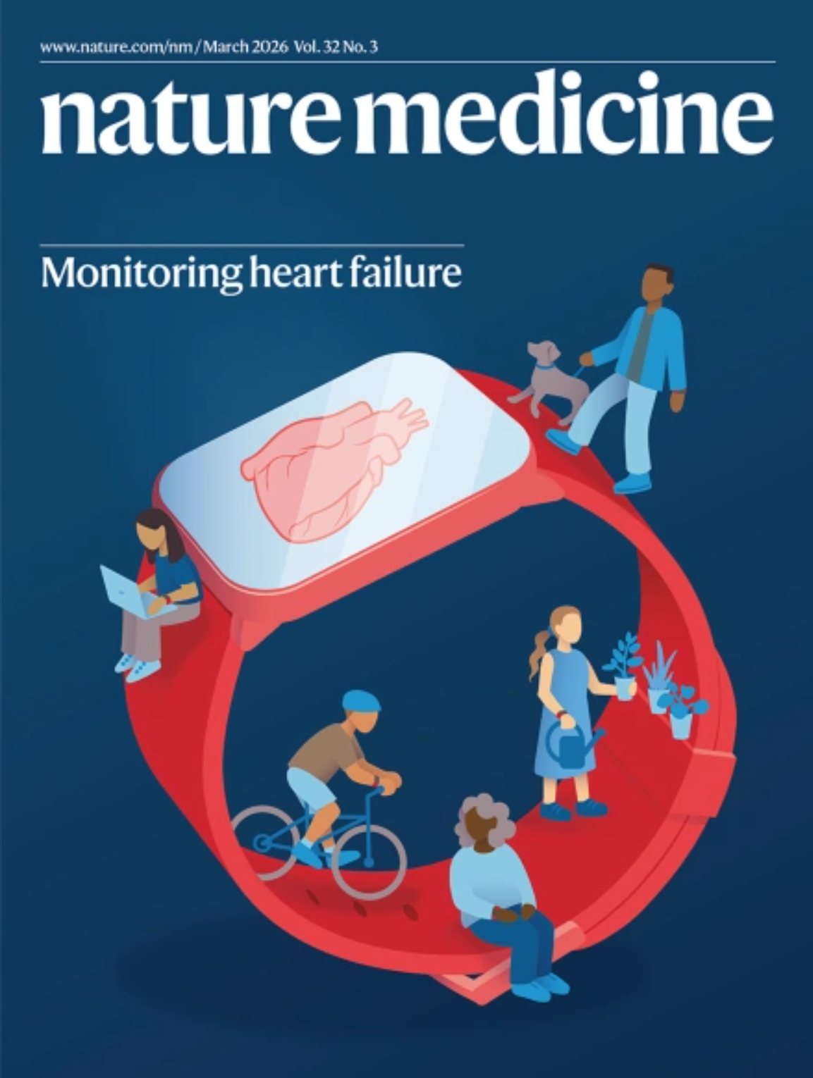
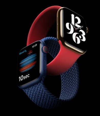
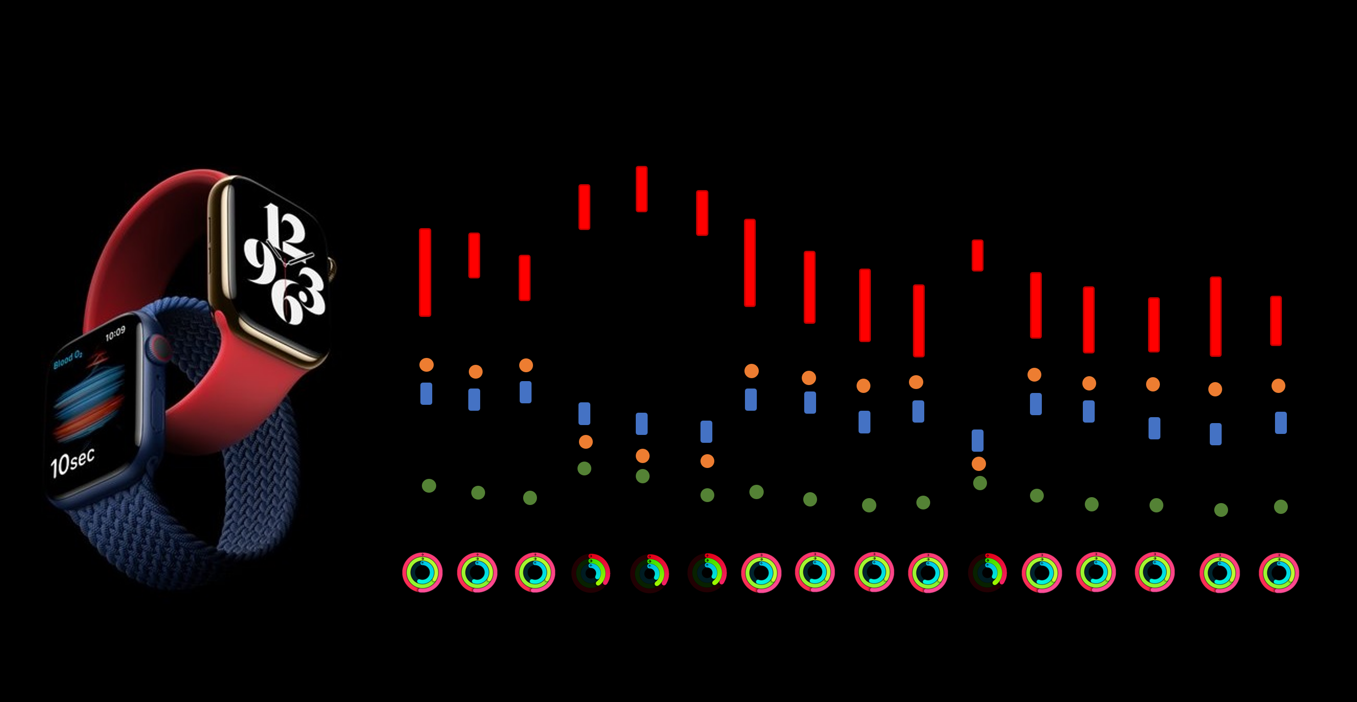
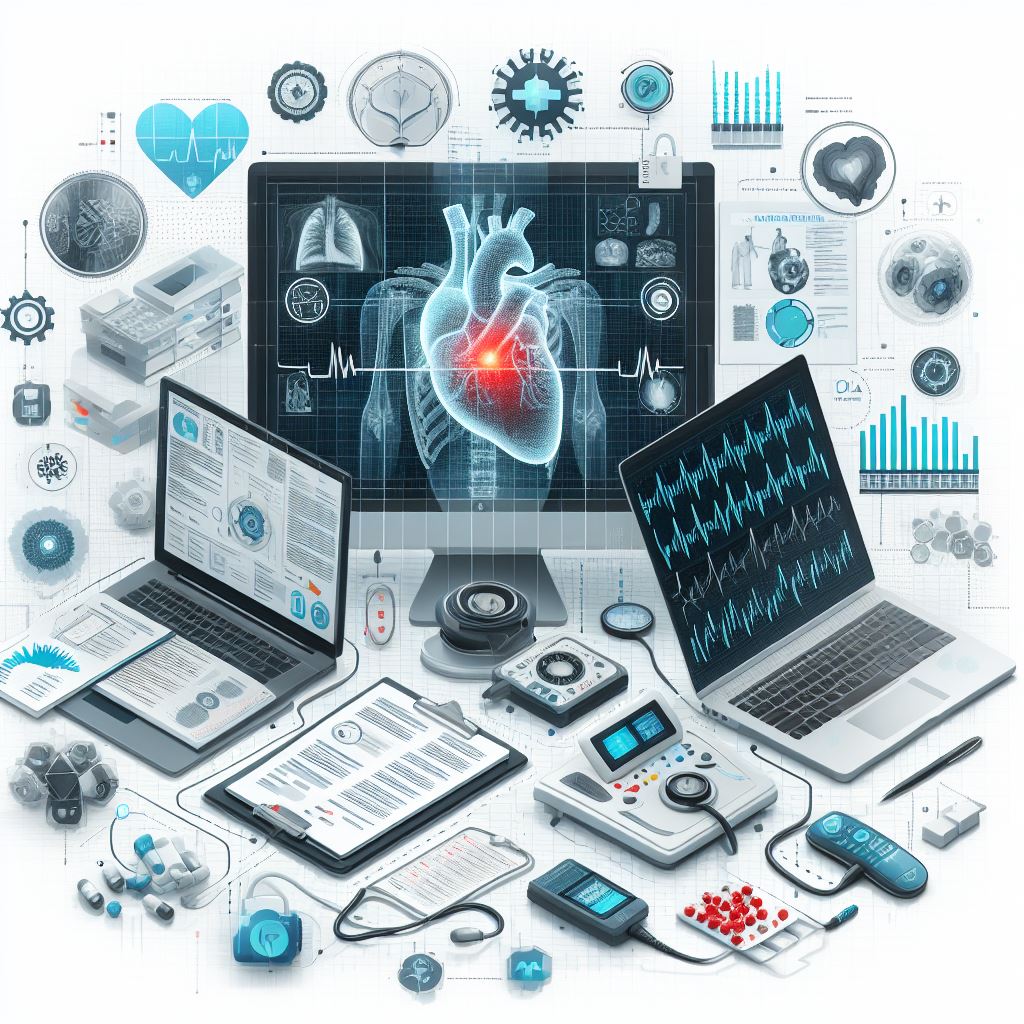
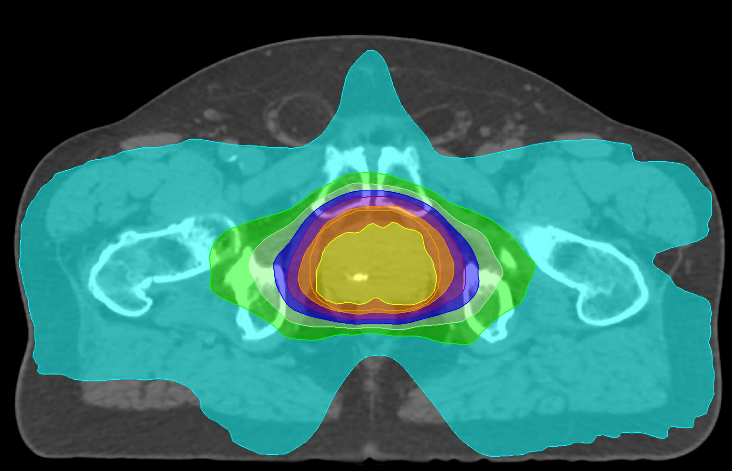
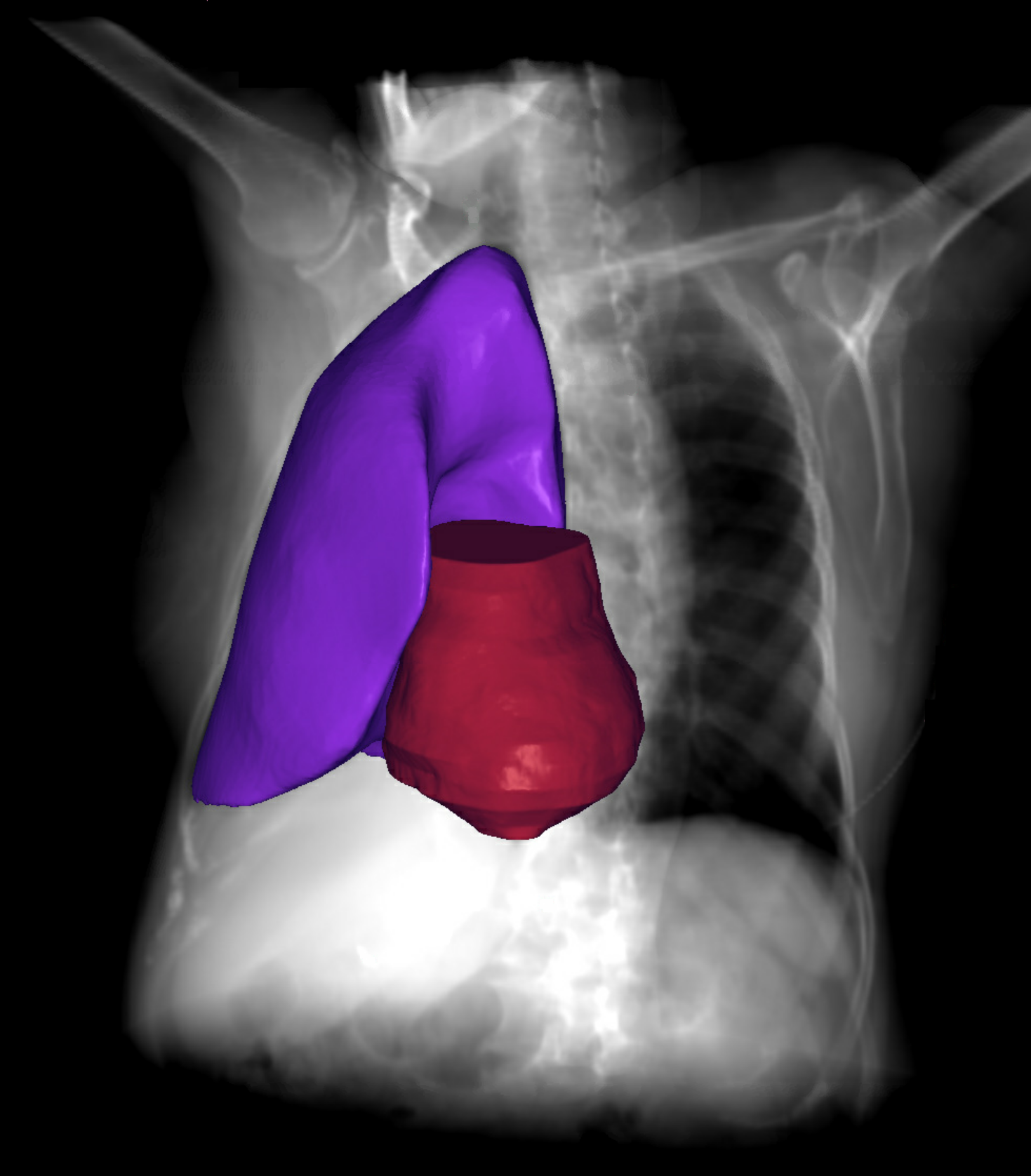
Medical domains: Our work includes a variety of data modalities (biological, wearable, structured reporting, and imaging) and critical diseases such as cancer and heart disease. We work closely with clinicians to understand and ultimately augment patient care with AI.

AI technologies: Solving clinical problems means pushing and extending the boundaries of AI technology including semi-supervised learning, domain adaptation/model generalization, meta-learning, and multi-modal data fusion.

Data: We work on datasets of all shapes and sizes, from rare diseases with rich structured data but few patients, to diverse imaging datasets in the tens of thousands.

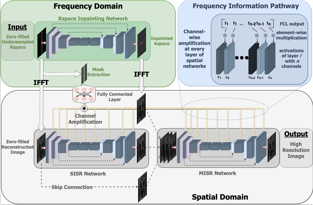
Keep reading below for example projects.

Wearable device biomarkers

What if we understood the patient journey instead of the Coles Notes?

Wearable technologies offer a unique glimpse into patient function and biology outside of episodes of care. If AI is the present, wearbles with AI are the next frontier. Working with leading cardiologists Heather Ross and Yas Moyaedi, the Ted Rogers Centre for Heart Research, and a key partnership with Apple we are developing AI technology to asses the role of wearbles in heart failure.

Multimodal Generative Models and Multitask Learning

What if a model could simultaneously understand multiple sources of medical data and multiple medical tasks?

Multimodal large language models in healthcare represent a cutting-edge fusion of advanced linguistic capabilities and diverse data sources. By seamlessly integrating text, images, electrocardiograms, and more these models harbor great potential to enhance clinical care through more robust and generalizable machine learning.

Automation in Radiation Therapy

The new standard of care

An experienced clinical team can more than double a cancer patient’s odds of survival. In particular, radiation therapy (RT) is a key part of the treatment for 40% of all cancer patients with the potential to be highly curative and effective given the right treatment plan. However, a major barrier in that effectiveness is the expertise and experience of the patient’s clinical team in his or her particular cancer. We believe that AI can help bring the expertise of the world’s best clinical teams to every patient in every hospital, saving lives and costly resources.

Working with clinical partners (Tom Purdie, Ale Berlin, and Leigh Conroy) we have developed the world’s first computer vision treatment system that we piloted to treat prostate cancer patients at Princess Margaret Cancer Centre, with superior results to our standard of care for the majority of patients. Cancer treatments are generated by the AI in minutes, instead of the hours-to-days taken under the typical manual process, increasing both efficiency and quality of care. Follow-up studies are expanding the models to breast cancer, brain tumours in young adults, and more. Through a successful licensing deal of this Canadian technology to RaySearch Laboratories we are also supporting deployment across Canada and around the world.

Image Segmentation

Coming soon to a clinic near you

Image segmentation is the gold standard for computational morphology and a key step in the radiation therapy process. A long sought after technology, automated segmentation methods are recently achieving near clinical performance paving the way for clinical studies, but many challenges remain.

Imaging Biomarkers

An untapped gold mine?

With global acquisition rates of medical imaging ever increasing, imaging represents a significant resource of potential prognostic value. Using combinations of deep learning and computer vision technologies to extract quantifiable features from imaging to risk stratify patients and better understand their care is growing area of research.

Jobs Help Wanted"Jobs Help Wanted" by Innov8social is licensed under CC BY 2.0

Do you want to change the shape of AI and healthcare?

  • Postdoctoral Fellowships

    We are seeking a postdoctoral fellow with experience in medicine and AI. Expertise is required in machine learning and programming in the context of at least one of medical signals, imaging, or wearable data.

    Interested students can submit a CV, a copy of your most relevant publication, and the names and contact information for up to three references to Chris McIntosh, with the subject line Postdoctoral application.

  • Graduate Students

    We are seeking motivated MSc/PhD students with a keen interest in medicine and AI. Expertise in at least one topic (machine learning, programming, and biology) and experience in others is desired.

    Interested students must apply to either the Department of Medical Biophysics (preferred), or Computer Science as appropriate to their interests and backgrounds.

  • Summer Students

    We welcome undergraduate internships/summer students with a keen interest in medicine and AI. Experience in at least one topic (machine learning, programming, and biology) is required.

    Interested students are encouraged to apply to the MBP summer student program.

Affiliations